| LIBROW | ® |
| Products | Solutions | Vehicles | Articles | Inquiry | Contacts | My account |
Article 12
3D median filter — ultrasound image despeckling
Limited offer
Professional Librow Calculatorvisit
for free
- Bessel functions
- gamma function
- complex numbers
7.4 MB for Windows

1. Introduction
Median filter is windowed filter of nonlinear class that is used for image quality improvement. In our basic article “Median filter” we have demonstrated capabilities of the median filter in “salt and pepper” noise reduction and in this case we show its ability to suppress speckle noise.
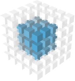
3D median filter uses 3D parallelepiped window to process volume image elements — like the one depicted in fig. 1.
 Fig. 1. 3D window of 3×3×3 size.
2. Speckle noise in ultrasound images
An inherent property of ultrasound imaging is the presence of speckle noise. Speckle is a random, deterministic, interference pattern in an image formed with coherent radiation of a medium containing many subresolution scatterers. The texture of the observed speckle pattern does not correspond to real structure. However, the local brightness of the speckle pattern does reflect the local echogenicity of the underlying scatterers.
Speckle has a negative impact on ultrasound image perception, reducing the image readability. This reduction in contrast resolution is responsible for the poorer effective resolution of ultrasound imaging compared to X-ray and magnetic resonance imaging (MRI).
3. Median filter — despeckle solution
To improve the image quality it is necessary to reduce speckle noise level. To solve the task median filter can be used. In this case for demonstration we are using weighted median filter with window of 3×3×3 size.
| a. |
|
b. |
|
In fig. 2a depicted the input 3D voxel volume — it is an original scanned ultrasound image — and fig. 2b shows the result of filtering. Click Play button to see heartbeat movie.
Pay attention to the level of speckle noise suppression. On the other hand since in our case median filter has non-adaptive nature one can see tissue information degradation as well — filter is blurring both noise component and useful information. To overcome this disadvantage adaptive feature preserving techniques can be used though they are usually much more complicated and not that straightforward as median filtering.
|
|
|||||||||||||
|
|||||||||||||


